
Early detection of esophageal, gastric and colorectal cancer with magnifying endoscopy and chromoendoscopy. Book a consultation with Dr. Younchi.
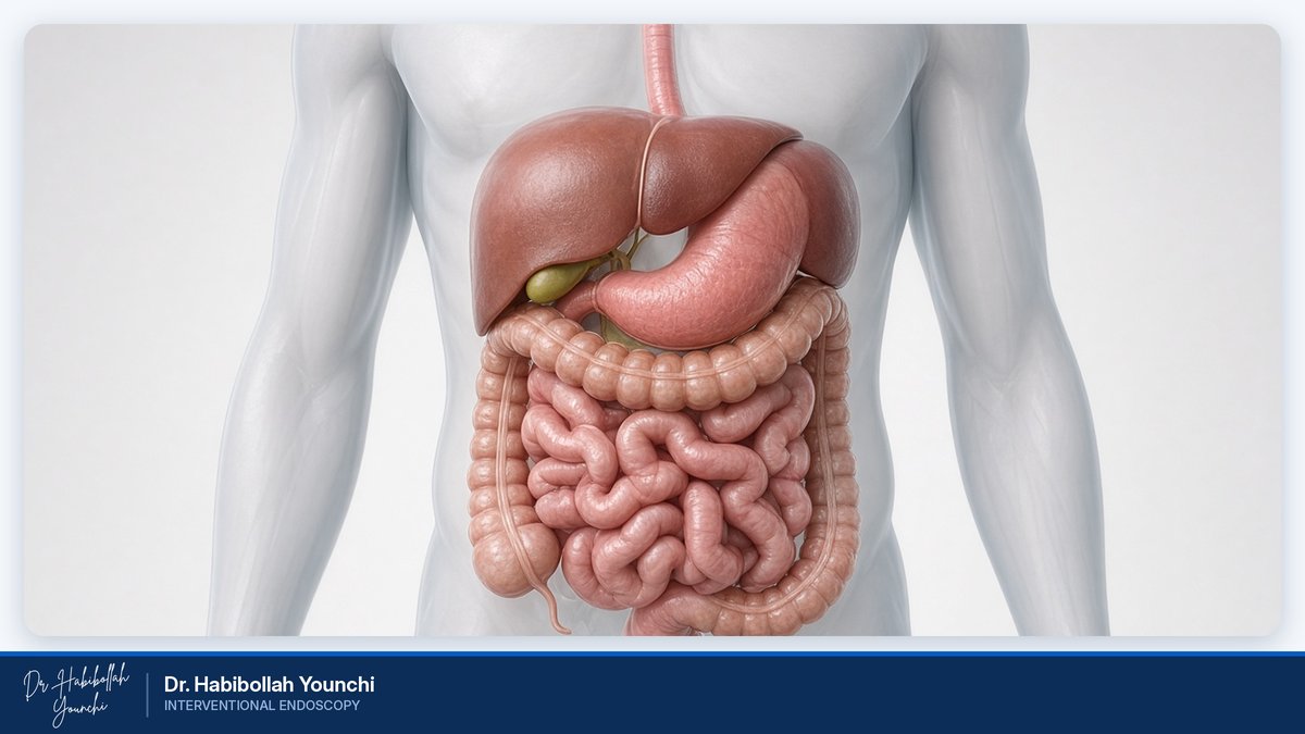
Early Detection of GI Cancer
What Is Early Gastrointestinal Cancer Detection?
Cancers of the esophagus, stomach and colon often develop slowly and silently. In their earliest stages, they may cause no symptoms at all. Advanced endoscopic imaging makes it possible to find early changes while they are still small, superficial and, in many cases, treatable without surgery. This page explains how early detection works, who should consider it, and what you can expect when you visit Dr. Habibollah Younchi, Consultant Internist and Interventional Endoscopist.
Early detection means identifying cancer, or the precancerous changes that can lead to it, before symptoms appear or while the disease is still confined to the inner lining of the digestive tract. Many gastrointestinal cancers begin as subtle changes in the mucosa: a slight change in color, a faint irregularity in surface texture, or an abnormal pattern of tiny blood vessels. Such findings are easy to miss during a routine examination.
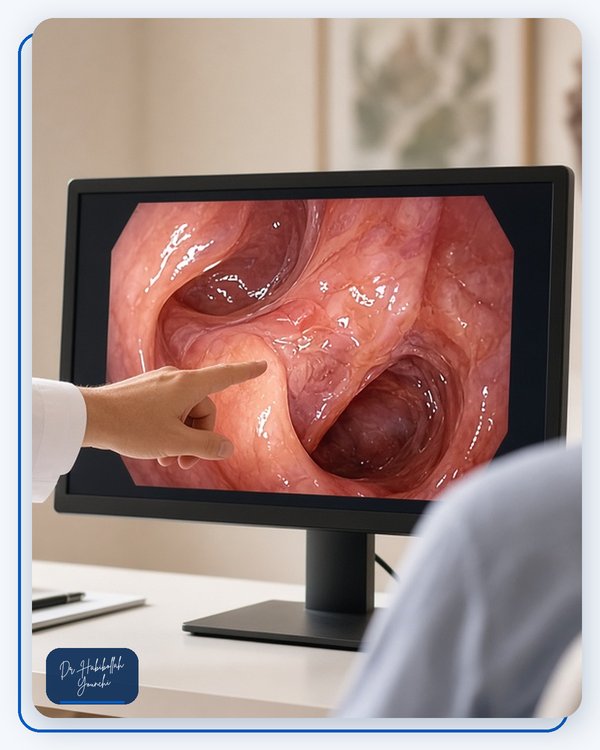
Modern endoscopy combines high-definition video, optical magnification and special contrast techniques to make such changes visible. When a suspicious area is found, a small tissue sample (biopsy) can be taken for laboratory analysis, giving a definitive diagnosis.
Why Early Detection Matters
- Better outcomes: Cancers found at an early stage are generally much more treatable than those diagnosed after symptoms have developed.
- Less invasive treatment: Many early lesions can be removed endoscopically, through the mouth or rectum, without open surgery and without removing the organ.
- Prevention, not just detection: Precancerous lesions, such as colon polyps or areas of dysplasia, can be removed before they have a chance to develop into cancer.
Who Should Consider Screening?
The right timing depends on your age, family history and personal risk factors. Consider an evaluation if any of the following apply to you:
- You are 45 or older. Many international guidelines now recommend that adults at average risk begin colorectal cancer screening at this age.
- You have a close relative who has had stomach, esophageal or colorectal cancer, or colorectal polyps.
- You have had a *Helicobacter pylori* infection, chronic gastritis, atrophic gastritis or intestinal metaplasia.
- You have long-standing heartburn or acid reflux, or have been diagnosed with Barrett's esophagus.
- You smoke, drink alcohol regularly, or habitually drink very hot beverages.
- You have a history of inflammatory bowel disease, such as ulcerative colitis or Crohn's disease.
Warning Signs You Should Not Ignore
Please see a doctor promptly if you notice any of the following:
- Difficulty or pain when swallowing
- Unintended weight loss
- Persistent indigestion, upper abdominal pain or feeling full after eating only a small amount
- Vomiting blood, or black, tar-like stools
- Blood in the stool or a lasting change in bowel habits
- Iron-deficiency anemia, persistent fatigue or unexplained shortness of breath
These symptoms are often caused by benign conditions, but they should always be evaluated.
Advanced Endoscopic Techniques
High-Definition and Magnifying Endoscopy
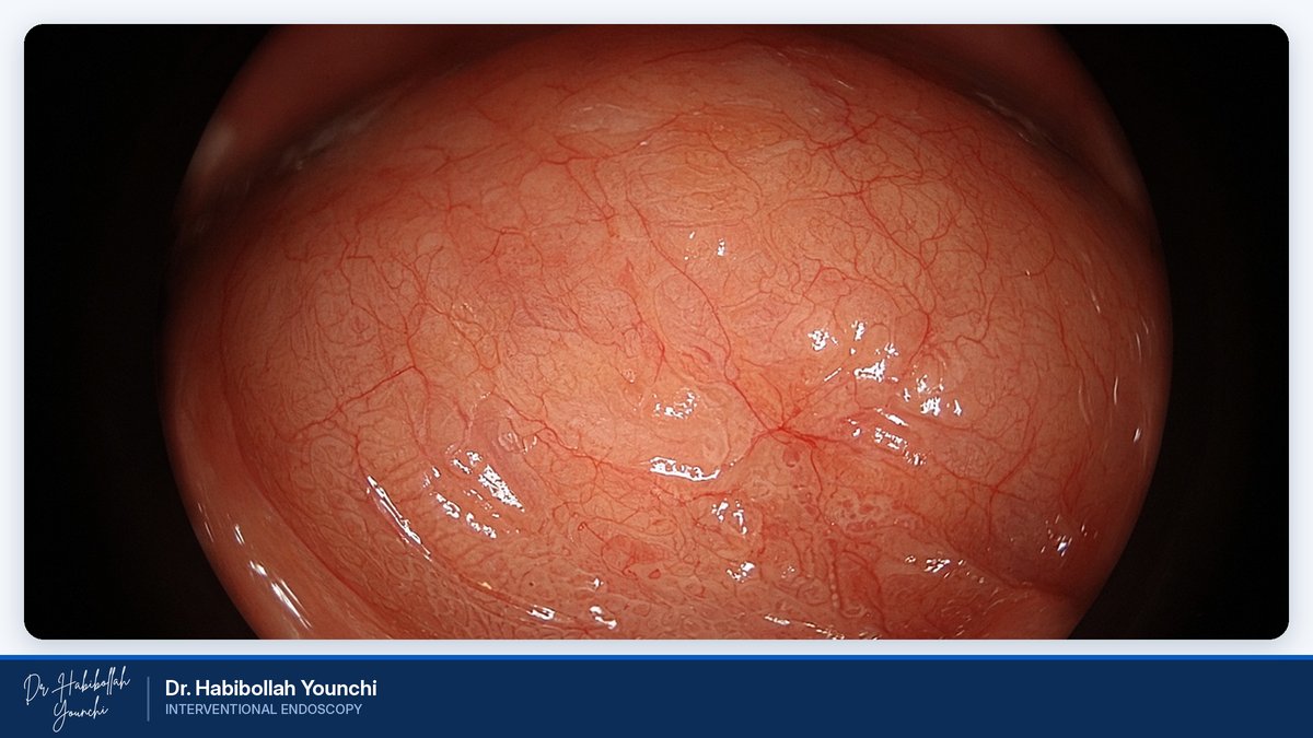
High-definition endoscopes provide sharp, detailed images of the digestive lining. Magnifying endoscopy goes a step further, enlarging the view optically so that the fine surface structure and the network of tiny blood vessels can be examined closely. Changes in these micro-patterns can help distinguish benign tissue from early neoplasia and can help define the borders of a lesion before treatment.
Chromoendoscopy
In chromoendoscopy, safe dyes or stains are sprayed onto the lining during the examination. Agents such as indigo carmine, Lugol's iodine and acetic acid highlight subtle irregularities that may not be visible under standard white light. Lugol's iodine, for example, is commonly used to evaluate the esophagus for early squamous lesions.
Image-Enhanced Endoscopy
Image-enhanced endoscopy, sometimes called virtual chromoendoscopy, uses filtered light or digital processing (for example, narrow-band imaging) to accentuate blood vessels and surface patterns without the need for dyes. Combined with magnification, it allows detailed, real-time assessment of suspicious areas.
Targeted Biopsy
When an area looks abnormal, small tissue samples are taken painlessly through the endoscope. Detailed imaging helps direct biopsies to the most relevant spots. A pathologist then examines the tissue to confirm the diagnosis and guide treatment.
Early Detection by Organ
Esophageal Cancer
The two main types of esophageal cancer are squamous cell carcinoma and adenocarcinoma. Squamous cell carcinoma is associated with smoking, alcohol and very hot beverages, while adenocarcinoma is linked to chronic acid reflux and Barrett's esophagus. Early lesions can be extremely subtle, which is why careful upper endoscopy, often supported by chromoendoscopy and magnification, is so valuable. Patients with Barrett's esophagus are usually advised to undergo regular surveillance.
Gastric (Stomach) Cancer
Gastric cancer is relatively common in Iran. It often develops through a sequence of changes, beginning with chronic inflammation, frequently caused by *Helicobacter pylori*, and progressing to atrophic gastritis, intestinal metaplasia and dysplasia. Detecting and treating *H. pylori*, identifying these precancerous conditions, and monitoring them appropriately are key steps in prevention. Magnifying and image-enhanced endoscopy can help reveal early gastric cancers that are flat or only slightly raised or depressed.
Colorectal Cancer
Most colorectal cancers develop from polyps over a period of years. Colonoscopy allows the doctor to find and remove polyps during the same procedure, which can reduce the risk of developing cancer. Stool-based tests are another screening option, but a positive result should be followed by colonoscopy. Advanced imaging helps characterize polyps and detect flat lesions that are harder to see.
What to Expect and How to Prepare
Before the procedure
- For an upper endoscopy, you will usually be asked not to eat or drink for several hours beforehand.
- For a colonoscopy, you will follow a special diet and take a bowel preparation solution so that the colon is completely clean. A well-prepared bowel is essential for an accurate examination.
- Tell your doctor about all of your medications, especially blood thinners, diabetes medications and iron supplements, as some may need to be adjusted.
- If you receive sedation, arrange for someone to accompany you home.
During the procedure
Most examinations are performed under sedation for your comfort. A thin, flexible endoscope is gently guided through the digestive tract while the doctor carefully inspects the lining, uses enhanced imaging when needed, and takes biopsies if required.
After the procedure
You will rest briefly while the sedation wears off. Mild bloating or a sore throat is common and usually settles quickly. Your doctor will explain the initial findings, and biopsy results are typically available within several days.
Treatment Options for Early Lesions
When a lesion is found early, it can often be treated endoscopically. The most appropriate option depends on the lesion's size, location, depth and appearance, as well as on the pathology results.
- Endoscopic mucosal resection (EMR): A polyp or superficial lesion is lifted away from the deeper layers, usually by injecting fluid beneath it, and then removed.
- Endoscopic submucosal dissection (ESD): Larger superficial lesions are removed in a single piece, which supports accurate pathological assessment.
- Full-thickness resection (FTRD): The Full Thickness Resection Device removes all layers of the gastrointestinal wall at the site of a lesion. It may be appropriate for carefully selected lesions that cannot be removed safely or completely with conventional techniques.
Not every lesion is suitable for endoscopic treatment. Where surgery or other therapy is the better option, you will be guided toward the most appropriate care.
Why Choose Dr. Habibollah Younchi?
- Over 28 years of medical experience: Dr. Younchi completed his specialty in internal medicine in 1997 and practices as a Consultant Internist and Interventional Endoscopist.
- A pioneer of magnifying endoscopy in Iran: He pioneered magnifying endoscopic imaging in Iran by introducing the first magnifying endoscopy device at Madaen Hospital, which he joined in 2008.
- First FTRD procedure in the region: He performed the region's first Full Thickness Resection Device (FTRD) procedure at Madaen Hospital.
- Extensive clinical experience: He has built a clinical database of more than 30,000 cases.
- Focused on early cancer detection: His work centers on detecting early gastric, esophageal and colon cancers using magnifying endoscopy and chromoendoscopy.
- Ongoing training: In 2023, he completed hands-on training in Endoscopic Sleeve Gastroplasty (ESG) in India.
Frequently Asked Questions
Is endoscopy painful?
Most patients experience little or no discomfort, as the procedure is usually performed under sedation. You may feel some bloating or have a mild sore throat afterward, which typically resolves within a day.
How is magnifying endoscopy different from a standard endoscopy?
Magnifying endoscopy enlarges the image of the digestive lining, allowing the doctor to study fine surface patterns and tiny blood vessels. This added detail can help identify and characterize early lesions that may be difficult to recognize with standard endoscopy alone.
I have no symptoms. Do I still need screening?
Possibly. Early gastrointestinal cancers and precancerous lesions often cause no symptoms. Whether you need screening depends on your age, family history and risk factors, so it is worth discussing with your doctor.
Are the dyes used in chromoendoscopy safe?
The agents used in chromoendoscopy are applied only to the surface of the digestive lining and are generally considered safe. Please tell your doctor about any allergies, especially to iodine, before the procedure.
If a lesion is found, will I need surgery?
Not necessarily. Many early lesions can be removed endoscopically. Your doctor will review the imaging and pathology findings with you and recommend the most suitable treatment for your situation.
How often should I be checked?
The interval depends on what is found during your examination and on your personal risk factors. After your procedure, you will receive a follow-up plan tailored to your results.
Book a Consultation
Dr. Habibollah Younchi is a Consultant Internist and Interventional Endoscopist with more than 28 years of experience. He introduced the first magnifying endoscopy device at Madaen Hospital and has dedicated his work to the early detection of gastric, esophageal and colon cancer using magnification, image-enhanced techniques and chromoendoscopy, supported by a clinical database of more than 30,000 cases.
If you would like to know whether image-enhanced endoscopy is right for you, please get in touch to arrange a consultation.
Phone: 02155958685
Mobile: 09391944012
Email: dryounchi@gmail.com
Instagram: dr.younchi
Please bring any previous endoscopy, colonoscopy or pathology reports with you.