
Magnifying endoscopy enlarges the lining of the digestive tract up to around 80 to 100 times, helping detect and characterize early changes in the esophagus, stomach and colon. Learn how it works, who may benefit and what to expect, with Dr. Habibollah Younchi, Consultant Internist and Interventional Endoscopist.
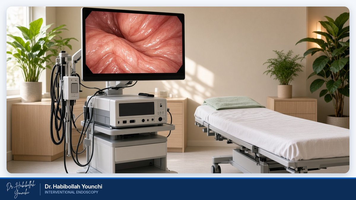
Magnifying Endoscopy for Early Detection of Digestive Tract Lesions
What Is Magnifying Endoscopy?
Magnifying endoscopy is an advanced form of endoscopy that allows the doctor to examine the inner lining of the digestive tract in far greater detail than a standard examination. Like a conventional endoscopy or colonoscopy, it uses a thin, flexible tube with a camera at its tip. The difference is that a magnifying endoscope contains a special optical zoom lens that can enlarge a selected area many times over, much like a microscope placed directly on the surface of the tissue.
This close-up view lets the endoscopist study features that are almost invisible at normal magnification: the fine structure of the lining's surface and the pattern of the tiny blood vessels beneath it. Changes in these patterns are often among the earliest signs that tissue is becoming abnormal.
Magnifying endoscopy does not replace a biopsy, which remains the standard for confirming a diagnosis. Instead, it helps the doctor decide where to take samples and how best to manage a suspicious area.
How Magnifying Endoscopy Works
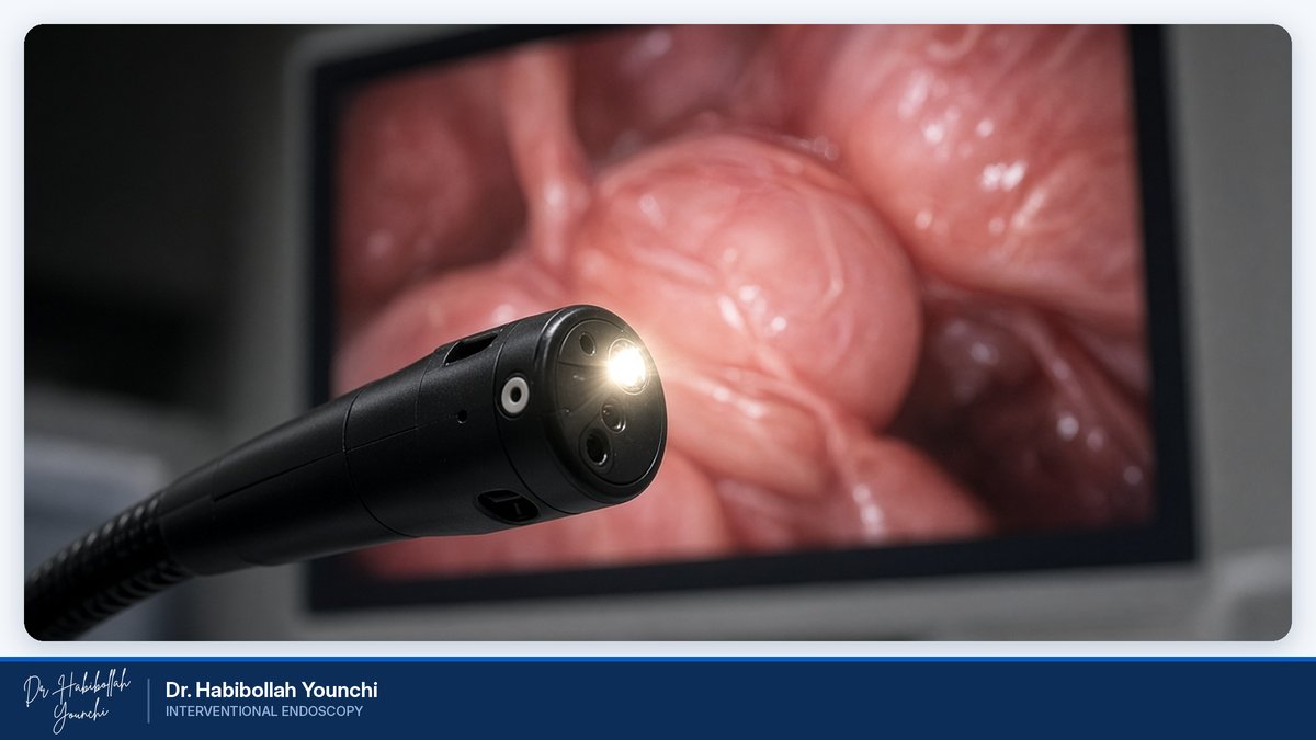
The examination begins like a routine endoscopy, with a careful survey of the lining in high-definition white light. When an area looks unusual, even very subtly, the endoscopist moves the tip of the instrument close to it and activates the optical zoom. Optical magnification enlarges the image without loss of sharpness, typically up to around 80 to 100 times, and even higher on some systems.
Magnification is usually combined with image-enhanced endoscopy. Narrow Band Imaging (NBI) and similar technologies from other manufacturers filter the light into specific wavelengths that are strongly absorbed by hemoglobin in the blood. As a result, small vessels stand out in sharp contrast against the surrounding lining.
Chromoendoscopy may also be used. In this technique, special dyes or solutions, such as indigo carmine, acetic acid or Lugol's iodine, are sprayed onto the surface to highlight its texture and borders.
Together, these tools allow the endoscopist to assess two key features. The first is the surface pattern, meaning the shape and arrangement of the tiny openings and ridges of the lining. The second is the vascular pattern, meaning the size, shape and distribution of the small blood vessels. Specialists interpret these findings using internationally recognized classification systems, which help distinguish normal or inflamed tissue from precancerous changes and early cancer.
Why It Matters: Key Benefits
Many early cancers and precancerous lesions of the digestive tract are flat, small and difficult to distinguish from normal tissue. Magnifying endoscopy offers several practical advantages in this setting.
Earlier recognition: Subtle abnormalities can be detected and examined before they cause symptoms.
Better characterization: The doctor can assess whether a lesion is more likely to be benign, precancerous or malignant.
Targeted biopsies: Samples can be taken from the most suspicious spot, which may improve diagnostic accuracy and reduce unnecessary sampling.
Treatment planning: The borders and depth of a lesion can be estimated more precisely, helping the doctor decide whether it may be suitable for removal through the endoscope rather than by surgery.
Same procedure, more information: For most patients, magnification simply adds detail to an examination they would already be having.
Conditions Magnifying Endoscopy Helps Evaluate
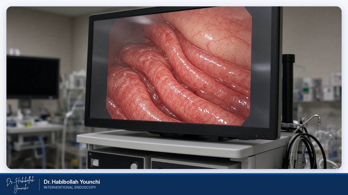
Magnifying endoscopy is most valuable for detecting and characterizing early, subtle changes in both the upper and lower digestive tract.
Early gastric cancer: In the stomach, magnification helps the endoscopist examine the fine surface and vessel patterns of suspicious areas. It is particularly useful in people with chronic gastritis, atrophic gastritis or intestinal metaplasia, conditions that are often associated with long-term Helicobacter pylori infection and that make early lesions harder to see.
Early esophageal cancer: Early squamous cell cancer of the esophagus can be very subtle. Magnifying endoscopy with NBI allows close study of the tiny looping blood vessels in the lining, whose shape and size change as tissue becomes abnormal. Lugol's iodine chromoendoscopy may be used alongside it.
Barrett's esophagus: Long-standing acid reflux can change the lining of the lower esophagus. Close inspection helps the doctor look for areas of dysplasia, which are precancerous changes that may need treatment or closer surveillance.
Colorectal polyps and lesions: During colonoscopy, magnification helps characterize polyps by their surface and vessel patterns. This can help predict whether a polyp is likely to be harmless, precancerous or cancerous, and guide the choice of removal technique.
Lesion margins: Magnification also helps define the margins of a lesion before endoscopic treatment.
Who May Benefit
Your doctor may recommend magnifying endoscopy if you have:
Family history: a close relative with stomach, esophageal or colorectal cancer.
Precancerous conditions: atrophic gastritis, intestinal metaplasia, Barrett's esophagus or a history of dysplasia.
Previous findings: a lesion or polyp found during an earlier endoscopy that needs closer assessment.
Persistent symptoms: such as difficulty swallowing, ongoing indigestion, unexplained anemia or bleeding.
Previous treatment: a need for follow-up after removal of an early cancer or a large polyp.
Other risk factors: such as long-standing reflux, smoking or regular alcohol use.
Not everyone needs magnifying endoscopy. The decision is made individually, based on your history, symptoms and previous results.
What to Expect and How to Prepare
Preparation is similar to that for a standard endoscopy or colonoscopy, and you will receive specific instructions before your appointment.
For an upper endoscopy, you will usually be asked not to eat or drink for about six to eight hours beforehand. For a colonoscopy, you will need to follow a special diet and take a bowel preparation solution so that the colon is clean enough for a detailed examination.
Please tell the medical team about all the medicines you take, particularly blood thinners, diabetes medicines and iron tablets, as some may need to be adjusted. Also mention any allergies, especially to iodine, and any heart, lung or kidney conditions.
On the day of the procedure, you will usually receive sedation to keep you relaxed and comfortable. Because the doctor takes time to examine suspicious areas closely, the procedure may take a little longer than a standard examination. Afterward, you will rest briefly while the sedation wears off. Please arrange for someone to accompany you home, and avoid driving for the rest of the day. Your doctor will explain the initial findings, and any biopsy results will follow once the laboratory analysis is complete.
Safety and Comfort
Magnifying endoscopy is performed with the same type of flexible instrument used for routine examinations and has a safety profile similar to that of a standard endoscopy. Endoscopy is generally a safe procedure, and serious complications are uncommon.
As with any medical procedure, some risks exist. These include reactions to sedation, bleeding after a biopsy or polyp removal, and, rarely, injury to the wall of the digestive tract. The dyes used in chromoendoscopy are applied only to the surface of the lining and are generally well tolerated. Your doctor will discuss the risks that apply to your particular situation before the procedure.
Most patients feel little or no discomfort during the examination. Afterward, you may notice mild bloating or a sore throat, which usually settles within a day. Please seek medical advice promptly if you develop severe abdominal pain, fever, black stools or bleeding.
Dr. Habibollah Younchi's Experience
Dr. Habibollah Younchi is a Consultant Internist and Interventional Endoscopist with more than 28 years of experience. He completed his specialty training in internal medicine in 1997 and has focused his career on advanced diagnostic and therapeutic endoscopy.
After joining Madaen Hospital in 2008, Dr. Younchi introduced the hospital's first magnifying endoscopy device. According to data from the leading endoscope manufacturers Olympus, Pentax and Fujifilm, he was a pioneer of magnifying endoscopic imaging in Iran.
His work centers on the early detection of gastric, esophageal and colon cancer, using magnifying endoscopy together with image-enhanced techniques and chromoendoscopy. Over the years, he has built a clinical database of more than 30,000 cases.
Dr. Younchi also performed the region's first procedure with the Full-Thickness Resection Device (FTRD), used to remove selected difficult lesions through the endoscope. In 2023, he completed training in Endoscopic Sleeve Gastroplasty (ESG) in India, reflecting his ongoing commitment to bringing modern endoscopic care to his patients.
Frequently Asked Questions
Q: How is magnifying endoscopy different from a standard endoscopy?
A: It uses the same type of flexible endoscope, but with an optical zoom lens that enlarges the image of the lining many times over. This allows the doctor to study fine surface and blood vessel patterns that are difficult to see during a standard examination.
Q: Is magnifying endoscopy painful?
A: Most patients feel little or no discomfort, as the procedure is usually performed under sedation. Mild bloating or a sore throat afterward is common and usually resolves within a day.
Q: Does it take longer than a regular endoscopy?
A: It may take a little longer, because the doctor spends extra time examining any suspicious areas in detail.
Q: Will I still need a biopsy?
A: Possibly. Magnifying endoscopy helps the doctor decide where to take samples, but a biopsy remains the standard way to confirm a diagnosis.
Q: Can magnifying endoscopy rule out cancer completely?
A: No test can guarantee that cancer is absent. Magnifying endoscopy provides highly detailed information that supports an accurate assessment, and your doctor will interpret it together with biopsy results and your medical history.
Q: What happens if an early lesion is found?
A: Your doctor will explain the findings and discuss the options with you. Depending on the type, size and depth of the lesion, it may be suitable for removal through the endoscope, or it may require further tests or treatment.
Book a Consultation
Dr. Habibollah Younchi is a Consultant Internist and Interventional Endoscopist with more than 28 years of experience. He introduced the first magnifying endoscopy device at Madaen Hospital and has dedicated his work to the early detection of gastric, esophageal and colon cancer using magnification, image-enhanced techniques and chromoendoscopy, supported by a clinical database of more than 30,000 cases.
If you would like to know whether image-enhanced endoscopy is right for you, please get in touch to arrange a consultation.
Phone: 02155958685
Mobile: 09391944012
Email: dryounchi@gmail.com
Instagram: dr.younchi
Please bring any previous endoscopy, colonoscopy or pathology reports with you.