
Image-enhanced endoscopy uses special light, dyes and digital processing to reveal subtle changes in the lining of the esophagus, stomach and colon. Learn about NBI, BLI, LCI, i-scan, chromoendoscopy and AI-assisted detection, the conditions they help find and what to expect, with Dr. Habibollah Younchi, Consultant Internist and Interventional Endoscopist.
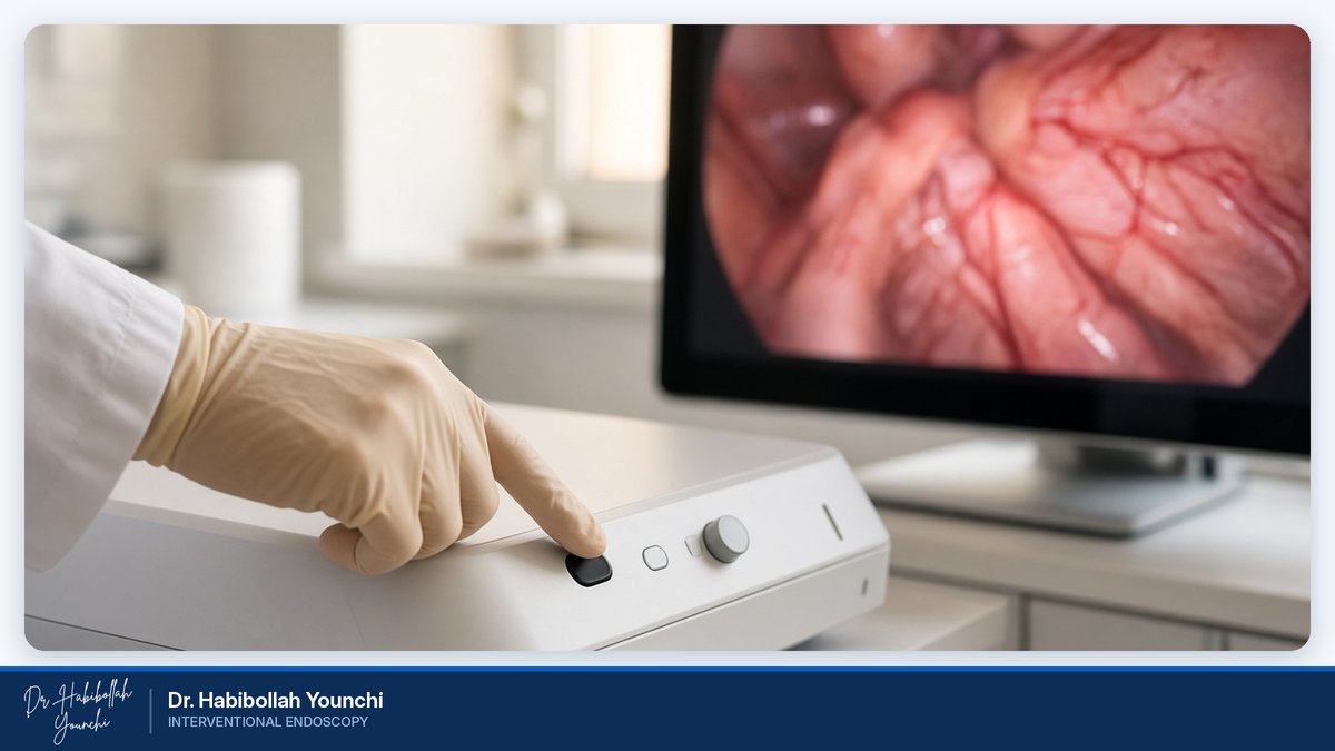
Image-Enhanced Endoscopy: Seeing Early Changes More Clearly
What Is Image-Enhanced Endoscopy?
Image-enhanced endoscopy, often shortened to IEE, is a group of techniques that make the lining of the digestive tract easier to read during an endoscopy or colonoscopy. A standard examination uses ordinary white light, which shows the lining much as the eye would see it. Many early lesions, however, are flat, pale or only slightly reddened, and they can blend into the healthy tissue around them.
Image-enhanced techniques change the way the lining is lit, colored or processed so that these subtle differences become visible. Some work by filtering the light, some by applying a harmless dye to the surface, and others by digitally refining the image in real time.
The aim is simple: to find small abnormalities sooner, to judge more accurately whether they are harmless or concerning, and to plan the most appropriate next step.
The Main Techniques
Several technologies fall under the umbrella of image-enhanced endoscopy, and an experienced endoscopist chooses among them according to the organ being examined and the question being asked.
Narrow Band Imaging (NBI) uses filtered blue and green light, which is strongly absorbed by hemoglobin. Blood vessels and the surface structure of the lining therefore appear in sharp contrast.
Blue Light Imaging (BLI) and Linked Color Imaging (LCI) are laser- and LED-based modes. BLI highlights vessels and surface detail in a similar way, while LCI enhances small differences in red tones, making inflammation and early lesions stand out against normal tissue.
i-scan is a digital enhancement system that adjusts sharpness, contrast and color tone at the press of a button.
Chromoendoscopy, the oldest of these methods, involves spraying a dye or solution onto the lining. Indigo carmine outlines the surface texture, acetic acid reveals fine patterns, and Lugol's iodine helps identify abnormal areas in the esophagus.
These modes can be combined with optical magnification for a closer look, and newer systems add computer-aided detection, in which artificial intelligence flags areas that deserve attention.
Benefits of Image-Enhanced Endoscopy
Image-enhanced endoscopy offers several practical advantages over white light alone.
It improves detection, helping the endoscopist notice flat or small lesions that might otherwise be overlooked. It supports characterization, giving valuable clues about whether a finding is likely to be benign or precancerous. It makes biopsies more targeted, so samples are taken from the most informative spots rather than at random. It defines the borders of a lesion, which is essential when planning endoscopic removal. Finally, it can help shape follow-up, as harmless findings may need less frequent surveillance.
None of these techniques replaces pathology, but together they make each examination more informative.
Conditions It Helps Detect
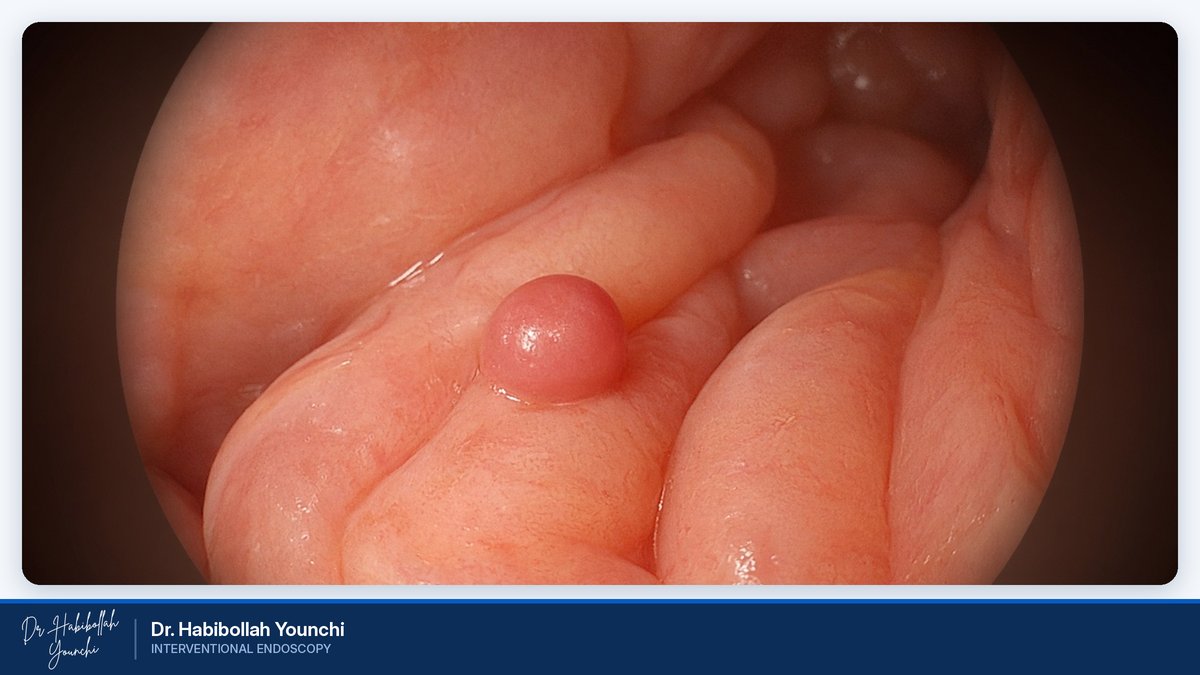
Image-enhanced endoscopy is valuable throughout the digestive tract, from the esophagus to the colon.
In the esophagus, it is widely used to examine Barrett's esophagus, a condition in which long-standing acid reflux changes the lining and raises the risk of cancer. Enhanced imaging helps locate areas of dysplasia, the precancerous changes that require treatment. It also assists in the early detection of squamous cell carcinoma, particularly in people with risk factors such as heavy smoking or alcohol use.
In the stomach, it helps identify intestinal metaplasia and atrophic gastritis, which are often related to long-term Helicobacter pylori infection, as well as early gastric cancer, which can be very subtle at its first stage.
In the colon, image-enhanced techniques improve the detection of adenomas and serrated polyps, both of which can develop into colorectal cancer if left in place. They also help predict the type of a polyp during the procedure, and they are used in surveillance colonoscopy for people with long-standing inflammatory bowel disease.
When a lesion is found early, it can often be removed endoscopically, without the need for surgery.
Who Should Consider It?
Image-enhanced techniques are used routinely during high-quality examinations, but they are especially important for certain patients.
You may benefit if you have Barrett's esophagus or long-standing reflux symptoms, a history of gastric intestinal metaplasia or atrophic gastritis, or a family history of gastric, esophageal or colorectal cancer. They are also helpful if you have had polyps removed before, if you live with ulcerative colitis or Crohn's disease affecting the colon, or if a previous examination showed an area that needs a closer look.
Your doctor will advise whether an enhanced examination is appropriate in your case.
The Procedure Step by Step
From the patient's point of view, an image-enhanced examination feels the same as a standard endoscopy or colonoscopy.
You will usually receive sedation so that you remain relaxed and comfortable. The endoscopist first inspects the lining in high-definition white light, then switches between enhancement modes, or applies a dye, wherever a closer assessment is needed. If a suspicious area is found, its pattern is evaluated and biopsies may be taken from the most relevant spot. In some cases, a small polyp can be removed during the same session.
The examination may take a few minutes longer than a routine one. Afterward, you will rest briefly while the sedation wears off, and the doctor will explain the initial findings.
Preparing for Your Examination
Good preparation is essential, because enhanced imaging is only as effective as the view allows.
For an upper endoscopy, you will normally need to stop eating and drinking for about six to eight hours beforehand. For a colonoscopy, you will receive instructions for a low-residue diet and a bowel-cleansing solution; a clean colon is critical for spotting small, flat lesions.
Tell your doctor about all your medications, especially blood thinners, antiplatelet drugs, diabetes medicines and iron tablets, as some may need to be adjusted. Please arrange for someone to accompany you home, since you should not drive for the rest of the day after sedation.
Safety
Image-enhanced endoscopy carries essentially the same low risks as a standard endoscopy or colonoscopy. Light-based modes such as NBI, BLI, LCI and i-scan simply change how the image is displayed and add no extra risk. The dyes used in chromoendoscopy are applied to the surface in small amounts and are generally well tolerated; patients with a known iodine allergy should mention it, as Lugol's iodine will then be avoided.
Mild bloating, a sore throat or temporary drowsiness are common and usually pass quickly. Serious complications, such as bleeding or perforation, are rare and are mainly associated with the removal of lesions rather than with the imaging itself.
Frequently Asked Questions
Is image-enhanced endoscopy a separate procedure?
No. It is performed during a standard endoscopy or colonoscopy, using the same instrument and the same preparation.
Can artificial intelligence replace the doctor?
No. Computer-aided detection acts as a second pair of eyes, but the endoscopist makes every decision about what a finding means and how to manage it.
Will the dye stain my body?
Chromoendoscopy dyes affect only the surface of the lining. Indigo carmine may give your stool a bluish or greenish color for a day, which is harmless.
Does enhanced imaging mean I will not need a biopsy?
Not necessarily. It guides where biopsies are taken, but pathology remains the standard for confirming a diagnosis.
How often should I have the examination?
This depends on your findings and risk factors. Your doctor will recommend an interval suited to your situation.
About Dr. Younchi and Booking an Appointment
Dr. Habibollah Younchi is a Consultant Internist and Interventional Endoscopist with more than 28 years of experience. He introduced the first magnifying endoscopy device at Madaen Hospital and has dedicated his work to the early detection of gastric, esophageal and colon cancer using magnification, image-enhanced techniques and chromoendoscopy, supported by a clinical database of more than 30,000 cases.
If you would like to know whether image-enhanced endoscopy is right for you, please get in touch to arrange a consultation.
Phone: 02155958685
Mobile: 09391944012
Email: dryounchi@gmail.com
Instagram: dr.younchi
Please bring any previous endoscopy, colonoscopy or pathology reports with you.